Foot and Ankle Anatomy
 It is a commonly used quote but Leonardo Da Vinci was correct when he stated that “The human foot is a masterpiece of engineering and a work of art”
This remarkably complex structure is made up of:
28 bones
19 muscles
107 ligaments
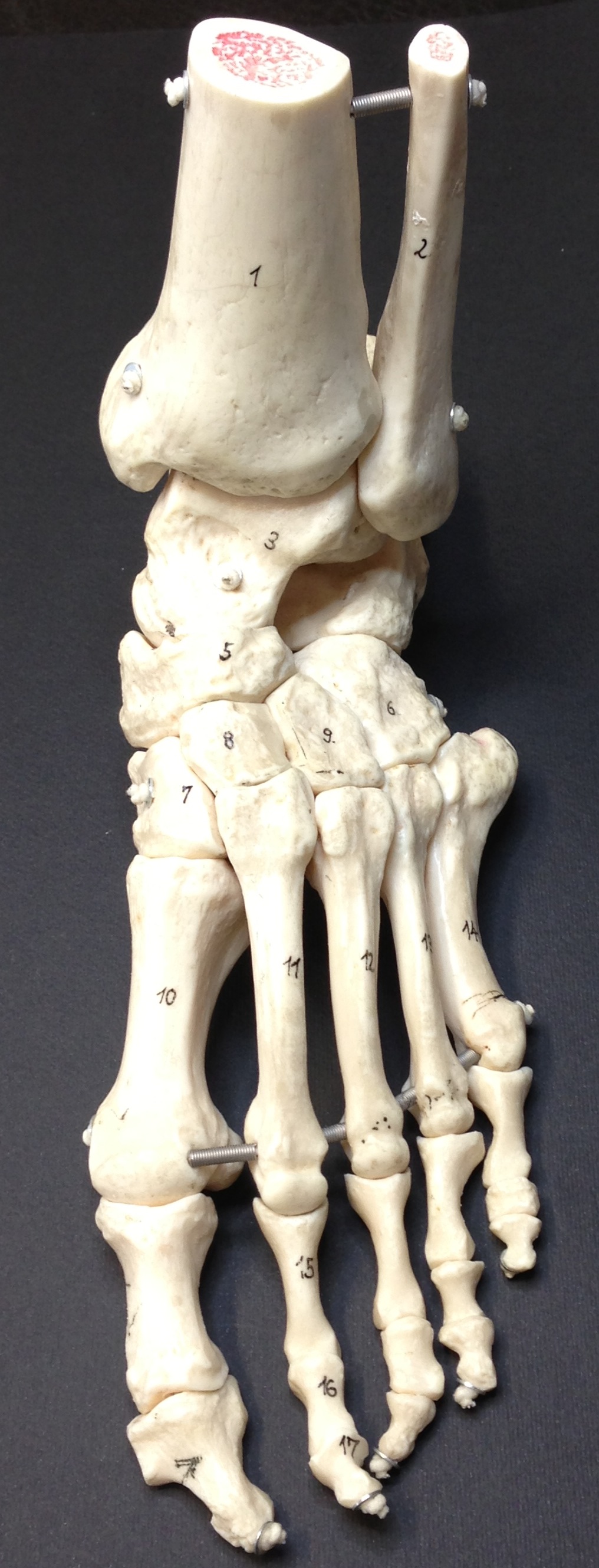
The foot is often divided into three segments, the rearfoot, midfoot and forefoot.
Rearfoot
This contains the largest bone in the foot – the calcaneus which articulates with the talus to form the subtalar joint that plays a significant role in foot function.
Midfoot
Contains the five tarsal bones, the cuboid on the outside of the foot, the navicular on the inside which articulates with the medial, intermediate and lateral cuneiform.
Forefoot
Consists of five metatarsals, 14 phalanges (toe bones) and two sesamoids located beneath the first metatarsal phalangeal joint (big toe joint)
The Ankle Joint
The talocrural joint or more commonly known as the ankle joint consists of three bones. The central pivot point the talus surrounded by the tibia and fibula, the joint is maintained on the inside by a very strong ligament called the deltoid ligament and on the outside by much weaker ligaments. One of these ligaments is the anterior talo-fibular ligament and is the most commonly injured ligament in the body. If you have ever twisted your ankle it is likely this ligament was injured.
The movements of the foot and ankle are intimately related; the subtalar and ankle joint interact closely to allow movement in three different directions that are fundamental to allow the foot and ankle to accept load and propel us forward:
- Up and down (plantarflexion and dorsiflexion)
- Left to right (abduction and adduction)
- Rotation left to right (inversion and eversion)